Introduction
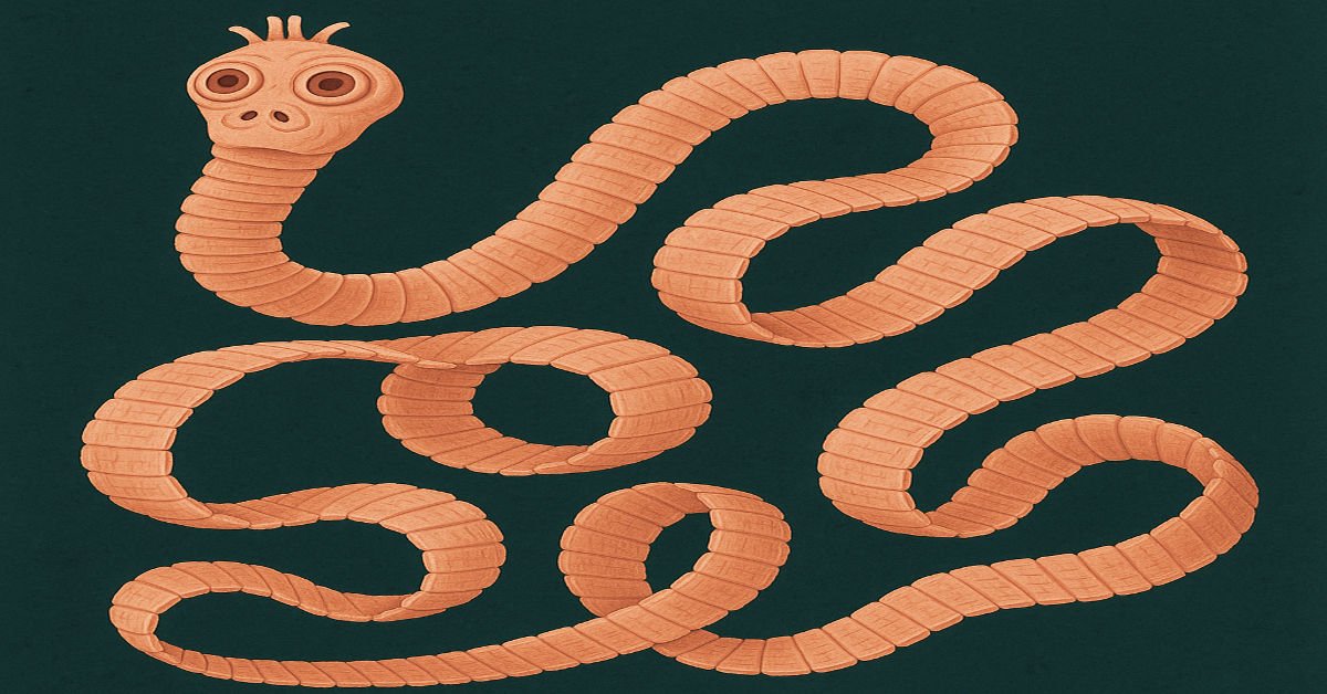
Taenia spp. are parasitic cestodes (tapeworms) that infect both animals and humans, leading to significant veterinary and public health concerns. These parasites primarily inhabit the small intestine of their final hosts while their larval stages (cysticerci) develop in the muscles and organs of intermediate hosts. Species such as Taenia saginata, Taenia solium, Taenia ovis, and Taenia hydatigena are commonly associated with infections in livestock and humans. Transmission occurs through the consumption of undercooked or contaminated meat, making meat inspection and proper cooking essential preventive measures. Effective diagnosis, treatment, and control strategies are crucial to reducing the economic and health burdens caused by these parasites.
Taxonomy and Pre-Dilection Site
| Category | Details |
|---|---|
| Family | Taeniidae |
| Genus | Taenia |
| Species | Taenia saginata, Taenia solium, Taenia ovis, Taenia hydatigena, T multiceps |
| Pre-Dilection Site | Small intestine (adult stage); muscles and organs (larval stage) |
Hosts
| Host Type | Examples |
| Final Host (F.H) | Humans (for T. saginata and T. solium), dogs, wild carnivores |
| Intermediate Host (I.H) | Cattle (T. saginata), pigs (T. solium), sheep (T. ovis), ruminants (T. hydatigena) |
Diagnosis and Differential Diagnosis of Taenia Infections in Animals
1. Diagnosis of Taenia Infections:
Taenia species are tapeworms that infect various animals, including dogs, cats, cattle, sheep, and pigs. Animals serve as either definitive or intermediate hosts depending on the species of Taenia. Diagnosis depends on the host species and the parasite’s life stage.
In Definitive Hosts (e.g., Dogs, Cats):
-
Fecal Examination: The Identification of Taenia eggs or proglottids (tapeworm segments) in feces is the most common method. Eggs are thick-shelled and can be seen under a microscope using flotation or sedimentation techniques.
-
Visual Observation: Owners may notice white, rice-like segments (proglottids) around the anus or in feces.
-
Molecular Tests: PCR testing can help distinguish Taenia species from other tapeworms, especially when eggs are morphologically similar.
In Intermediate Hosts (e.g., Cattle, Sheep, Pigs):
-
Post-Mortem Inspection: In livestock, cysts (like Cysticercus bovis or Cysticercus cellulosae) can be found in muscles or organs during meat inspection.
-
Imaging: In rare cases, ultrasound or radiographs may detect cysts in soft tissues.
-
Serology: Blood tests may detect antibodies in animals exposed to the larval stage, though this is not always routine.
2. Differential Diagnosis:
Several parasitic and non-parasitic conditions can mimic Taenia infections, especially when based on clinical signs or cyst presence.
For Adult Tapeworm Infection (Definitive Host):
-
Dipylidium caninum: This is also a tapeworm in dogs and cats; eggs are usually in packets, and the intermediate host is the flea.
-
Echinococcus spp.: Morphologically similar eggs; zoonotic and of public health concern. Requires careful species identification.
-
Spirometra spp. (Zipper tapeworms): Occurs in dogs and cats but has a different life cycle and egg morphology.
For Cystic Forms (Intermediate Hosts):
-
Hydatid Disease (Echinococcus): Forms large fluid-filled cysts, often in the liver and lungs; must be distinguished from Taenia cysts.
-
Sarcocystis spp.: Causes cysts in muscle tissues of livestock, but are microscopic and have different histological features.
-
Toxoplasma gondii: Can form tissue cysts, but generally smaller and often found in cats and intermediate hosts.
-
Abscesses or Tumors: In livestock, large cysts may resemble abscesses or neoplastic growths on meat inspection.
Pathology and Clinical Signs
The pathology of Taenia infections varies depending on the stage and species of the parasite:
| Type | Description | Clinical Signs |
| Intestinal Taeniasis | Caused by adult Taenia spp. in the intestines of final hosts. | Mild to no symptoms, occasional digestive discomfort. |
| Cysticercosis | Larval stage (Cysticercus) infection in muscles and organs of intermediate hosts. | Muscle weakness, and seizures (if neurocysticercosis in T. solium). |
| Coenurosis | The larval stage (Coenurus) of T. multiceps in ruminants and humans. | Neurological signs, ataxia, blindness. |
Life Cycle of Taenia spp.
Taenia species are parasitic tapeworms (cestodes) that infect various mammals, such as livestock, wild animals, and humans. Below is an original, plagiarism-free explanation of the Taenia spp. Life cycle in animals.
1. Adult Tapeworm in Definitive Host
-
Host: Carnivorous or omnivorous mammals (e.g., dogs for Taenia pisiformis, humans or pigs for Taenia solium).
-
Location: The mature tapeworm resides in the small intestine, anchoring itself with a scolex featuring suckers or hooks.
-
Reproduction: The worm produces segments called proglottids, each packed with numerous eggs. These proglottids break off and exit the host through feces.
2. Egg Spread in the Environment
-
Survival: The eggs are robust, capable of enduring harsh environmental conditions for weeks or months, increasing the chance of reaching an intermediate host.
3. Ingestion by Intermediate Host
-
Host: Herbivorous or omnivorous animals (e.g., cattle for Taenia saginata, rabbits for Taenia pisiformis).
-
Larval Release: Inside the host’s gut, eggs hatch into oncospheres (larvae), which burrow through the intestinal wall and enter the bloodstream.
4. Larval Development in Tissues
-
Migration: Oncospheres circulate via the bloodstream to organs or tissues, such as muscles or the liver.
-
Cyst Formation: Larvae transform into cysticerci, encapsulated larval stages that remain dormant but infectious. For instance:
-
Taenia solium forms cysts in pig muscles.
-
Taenia saginata forms cysts in cattle tissues.
-
-
Persistence: Cysticerci can survive for months, awaiting consumption by a definitive host.
5. Infection of Definitive Host
-
Transmission: A definitive host ingests raw or undercooked meat containing viable cysticerci.
-
Development: In the host’s intestine, cysticerci attach and mature into adult tapeworms, restarting the cycle as they produce egg-filled proglottids.
| Stage | Description |
| Egg Stage | Eggs are shed in the feces of infected final hosts. |
| Ingestion by Intermediate Host | Eggs hatch in the intestines and release oncospheres. |
| Larval Development | Larvae penetrate tissues and form cysticerci (cysts). |
| Ingestion by Final Host | Cysticerci develop into adult tapeworms upon consumption of infected tissue. |
| Maturation and Egg Production | Adults reside in the intestines and produce eggs, continuing the cycle. |
Epidemiology and Transmission
- Taenia infections have a worldwide distribution, with higher prevalence in areas practicing raw or undercooked meat consumption.
- Infection is common in livestock-rearing regions with poor meat inspection and hygiene.
- Human cysticercosis occurs due to the fecal-oral transmission of T. solium eggs, leading to severe neurological disorders.
Treatment and Control
- Medications to Eliminate Parasites:
- Praziquantel: Widely used, typically a single 5-10 mg/kg dose for Taenia saginata or T. solium. It targets adult worms in the gut.
- Niclosamide: Another option, given as a 2 g single dose for adults (chewed well). It works locally in the intestines.
- Albendazole: Preferred for cysticercosis (larval T. solium infection), dosed at 15 mg/kg/day for 8-30 days, often paired with steroids to control swelling.
- Managing Cysticercosis:
- For neurocysticercosis (brain involvement), antiparasitic drugs like albendazole or praziquantel are used cautiously with corticosteroids (e.g., prednisone) to reduce inflammation. Surgery may be required for large cysts or complications like fluid buildup in the brain.
- If cysts are inactive or asymptomatic, treatment may focus on observation rather than drugs.
- Additional Care:
- Seizure medications (e.g., levetiracetam or phenytoin) for neurocysticercosis-related seizures.
- Pain relievers or anti-inflammatory drugs for symptom relief.
- Surgical intervention for rare cases like intestinal blockages or brain cysts causing pressure.
- Monitoring:
- Follow-up stool tests 1-3 months after treatment to ensure the parasite is gone.
- Brain imaging (MRI/CT) to track cyst resolution in neurocysticercosis cases.
Control Strategies for Taenia:
- Hygiene Practices:
- Wash hands thoroughly after bathroom use and before cooking or eating.
- Avoid eating undercooked or raw beef (T. saginata) or pork (T. solium).
- Safe Food Handling:
- Freeze meat at -20°C for at least 7 days if not cooking immediately.
- Clean vegetables carefully to remove any tapeworm eggs from contaminated soil.
- Environmental Sanitation:
- Ensure proper disposal of human waste to prevent contamination of water or farmland.
- Build and maintain effective sewage systems to limit T. solium egg spread.
- Animal Husbandry: .
- Regular veterinary checks and deworming for livestock to reduce infection rates.
- Community-Based Prevention:
- Implement large-scale deworming programs in high-risk areas, using drugs like praziquantel for humans and oxfendazole for pigs.
- Educate communities about safe cooking, hygiene, and tapeworm risks.
- Use pig vaccines (e.g., TSOL18) where available to prevent T. solium infection.
- Early Detection and Treatment:
- Test and treat infected individuals to stop transmission, especially in endemic regions.
- Screen household contacts of T. solium cases, as eggs can spread from person to person.
Summary
Treatment
| Drug | Effectiveness |
| Praziquantel | Highly effective against adult Taenia tapeworms. |
| Albendazole | Used for cysticercosis and larval stages. |
| Niclosamide | Alternative treatment for adult cestodes. |
Control Measures
| Control Method | Description |
| Meat Inspection | Prevent the consumption of infected meat with cysticerci. |
| Proper Cooking | Cooking meat thoroughly kills cysticerci. |
| Sanitation and Hygiene | Proper waste disposal to prevent egg contamination. |
| Deworming Programs | Regular treatment of livestock and dogs to reduce transmission. |
Economic and Public Health Impact
- Taenia infections cause economic losses due to meat condemnation, reduced livestock productivity, and veterinary expenses.
- Neurocysticercosis, caused by T. solium, is a major cause of epilepsy in endemic areas, posing significant public health concerns.
Conclusion
Taenia spp. are important zoonotic cestodes that affect both animals and humans. Effective diagnosis, treatment, and control measures are essential to mitigate their impact. Strict meat inspection, proper hygiene, and regular deworming programs are key to reducing infections.
FAQs
1. What are the common hosts for Taenia spp.?
Taenia species commonly infect humans, dogs, and wild carnivores as final hosts, while cattle, pigs, and ruminants serve as intermediate hosts.
2. How is Taenia diagnosed in animals?
Diagnosis is based on fecal examination, serological tests, meat inspection, and post-mortem examinations to detect cysticerci or adult cestodes.
3. What are the symptoms of Taenia infection in animals?
Intermediate hosts may show muscle weakness, weight loss, or neurological symptoms in severe cases, while final hosts usually exhibit mild digestive discomfort.
4. How can Taenia infections be prevented?
Preventative measures include proper meat inspection, thorough cooking, maintaining good sanitation, and implementing deworming programs.
5. What is the impact of Taenia infections on public health?
Taenia solium causes neurocysticercosis in humans, a serious neurological disease leading to epilepsy and other severe complications.

