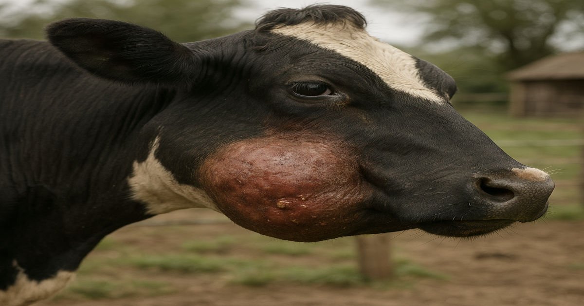
Actinomycosis disease, also known as Lumpy jaw, is sporadic but common in cattle. Caused by Actinomyces bovis, it usually develops after mouth injuries, leading to firm swellings and tissue damage. Although not contagious, it hampers feeding and overall health. Prompt diagnosis and care are crucial to limit complications.
Epidemiology of Actinomycosis:
-
The disease is sporadic but commonly seen in cattle.
-
Other animals—such as pigs, horses, goats, and dogs—can be affected sporadically, with human cases being exceptionally rare.
-
Despite its irregular occurrence, it is globally distributed and poses a concern due to poor treatment response.
-
Actinomyces bovis is a normal resident of the bovine mouth, becoming infectious after tissue injury.
-
Oral wounds from sharp feed or foreign bodies allow bacterial entry.
-
Erupting teeth in young cattle increase their susceptibility to infection.
-
Infections in the digestive tract may result from the ingestion of sharp materials that damage the mucosal lining.
Pathogenesis of Actinomycosis:
-
The infection begins when sharp feed particles or foreign objects injure the oral mucosa or gums, allowing Actinomyces bovis to enter.
-
Once inside, the bacteria trigger inflammation, leading to periostitis and osteomyelitis, particularly in jawbones.
-
This results in a pyogranulomatous osteomyelitis, where bone tissue is gradually destroyed.
-
Affected animals suffer mechanical difficulties in chewing (mastication) and grasping food (prehension).
-
If the digestive tract is involved, it may interfere with rumen function and digestion, contributing to weight loss and malnutrition.
-
In rare cases, the bacteria spread via the bloodstream, infecting other internal organs.
Clinical Findings of Actinomycosis:
In Cattle
-
Early Signs: Begins as a painless, hard swelling on the jawbones (mandible or maxilla), often near the molars.
-
Swelling Characteristics:
-
May be diffuse or localized.
-
Commonly thickens the lower jaw or shows in the space between the jawbones.
-
Often unnoticed until treatment is no longer effective.
-
-
Lesion Progression:
-
Can grow quickly (weeks) or slowly (months).
-
Eventually becomes painful, immobile, and may rupture the skin, forming draining sinuses.
-
Discharge contains sticky pus with small yellow-white granules.
-
Old sinuses may heal while new ones develop.
-
-
Effects on Eating:
-
Misaligned or painful teeth due to bone damage.
-
Leads to difficulty chewing, weight loss, and poor body condition.
-
-
Severe Complications:
-
Infection may spread to muscles and throat tissues, causing difficulty breathing.
-
No lymph node involvement.
-
In advanced cases, animals may become severely emaciated, requiring euthanasia.
-
-
Soft Tissue Involvement:
-
Commonly affects the esophageal groove, lower esophagus, and reticulum.
-
Leads to digestive problems: diarrhea, chronic bloat, and abnormal eating behavior (allotriophagia).
-
Rare cases involve testicles (orchitis), trachea (causing breathing issues), brain, or lungs.
-
In Pigs
-
Actinomycosis is rare but may cause:
-
Wasting due to internal organ lesions.
-
Occasionally, large granulomatous skin lesions or involvement of the udder.
-
Clinical Pathology of Actinomycosis:
-
Pus Smears:
-
Discharging pus is stained using the Gram stain.
-
This is a quick and effective way to confirm the diagnosis.
-
-
Non-Draining Lesions:
-
If pus is not discharging, diagnosis can be made using:
-
Tissue biopsies from the lesion core.
-
Aspirated fluid from the swelling.
-
-
Yellow granules (also called sulfur granules) from pus, when crushed and stained, help identify the bacteria microscopically.
-
Necropsy Findings – Actinomycosis in Cattle:
During necropsy, the most common observations include bone rarefaction and the formation of small cavities (loculi) and sinus tracts filled with thin, whey-like pus containing gritty granules. A consistent feature is the presence of dense fibrous tissue surrounding these lesions. The infection may spread to nearby soft tissues.
A hallmark of the disease is the presence of “club” colonies—structures made up of filamentous bacteria, which are visible under a microscope when examining smears from the pus granules or histological tissue sections.
Granulomatous abscesses filled with pus may also be detected in the esophageal groove, lower esophagus, and the front part of the reticulum. From these locations, the infection can extend into the surrounding area, leading to chronic localized peritonitis.
Digestive disturbances may be evident, such as unusually loose rumen contents, an empty abomasum, and mild inflammation of both the abomasum and intestines (abomasitis and enteritis). Notably, lymph nodes near the affected areas typically remain unaffected, regardless of the primary lesion site.
Treatment and Control of Actinomycosis in Cattle:
Treatment:
-
Iodide Therapy:
The most common approach involves using iodides either orally or intravenously.-
Intravenous Sodium Iodide: A 10–20% solution is administered slowly at a dose of 70 mg/kg body weight. This may be repeated after 1–2 weeks.
-
Oral Potassium Iodide: Given at 6–10 grams per animal daily for at least 10 days. Success varies, and much of the evidence is anecdotal.
-
-
Isoniazid Therapy:
Administered orally at 10–20 mg/kg body weight daily for around 30 days. It may stop lesion growth, but advanced cases often show poor response. -
Surgical Debridement:
Involves the removal of affected tissue and is often combined with drug therapy for better results. -
Cryotherapy:
Repeated application of liquid nitrogen has been reported as helpful in reducing lesions. -
Antibiotic Therapy:
In severe or non-responsive cases, antibiotics like penicillin, ampicillin, tetracyclines, or florfenicol may be used through injection.
Control Measures:
-
Isolation or Removal:
Animals with discharging lesions should be isolated or culled to prevent environmental contamination. -
Environmental Management:
While the disease is not highly contagious, it may spread if there are predisposing factors like oral injuries from rough feed. Proper feeding and management practices help reduce risk.
Conclusion:
Actinomycosis in animals, especially in cattle, is a chronic and progressive bacterial infection primarily affecting the bones and soft tissues of the head. Characterized by firm swellings, draining sinuses, and the presence of sulfur granules, this condition can severely impact the animal’s ability to eat and thrive. Although the disease is not highly contagious, it requires timely diagnosis and appropriate treatment, usually involving surgical drainage and long-term antibiotic or iodide therapy. Early intervention not only improves recovery outcomes but also prevents complications and economic losses in livestock management. Preventive strategies such as avoiding coarse feed and maintaining oral health play a key role in reducing the risk of infection.
Frequently Asked Questions (FAQs)
Q1. What animals are most commonly affected by actinomycosis?
A: Cattle are the most frequently affected species, but the disease can also occur in other domestic animals like pigs, horses, and, occasionally, sheep and dogs.
Q2. What causes actinomycosis in animals?
A: Actinomycosis is primarily caused by Actinomyces bovis, a bacterium that enters the body through mucosal wounds in the mouth, often due to rough feed or dental issues.
Q3. What are the clinical signs of actinomycosis?
A: Common signs include hard swellings on the jaw (commonly referred to as “lumpy jaw”), draining sinuses, difficulty chewing, and the presence of thick pus containing granules.
Q4. How is actinomycosis diagnosed?
A: Diagnosis is typically based on clinical signs, microscopic examination of pus for sulfur granules, radiography, and sometimes bacterial culture or biopsy.
Q5. Is actinomycosis contagious among animals?
A: No, actinomycosis is not considered contagious. It occurs sporadically when the bacteria gain entry through local trauma.